An article from The Academy of Finland
Simulation models to determine health of skeletal system
Researchers in Finland are using technical simulation models to simulate the functions of the human body. The method could help in the optimisation of exercise equipment for bones in the hip area.
Modelling is commonly used to simulate the operation of various types of machinery and equipment, and technical simulation models are also used to teach people how to use machines.
Now these models are being put to use in simulating the functions of the human body.
In an effort to combine their respective areas of expertise, the Lappeenranta University of Technology (LUT), the University of Jyväskylä and the UKK Institute for Health Promotion Research have joined forces to study how different exercises induce bone strain and strain rates.
“The idea to use simulation in studies with humans came completely by chance,” says Aki Mikkola, professor of virtual design at LUT.
“We were receiving inquiries from the Faculty of Sport and Health Sciences at Jyväskylä asking whether it would be possible to measure different aspects of human function. From a mechanical engineering point of view, none of this was a problem. Eventually, the inquiries became so frequent that we decided to go and see for ourselves what exactly the problem was.”
Computer modelling of human function
Simulation is based on well-known laws of physics. Computers are put to work to solve complex equations with a view to gaining simultaneous control over large and complicated systems.
“The simulation technique was originally developed to simulate the operation of combine harvesters and other mobile heavy duty machines,” explains Mikkola. “A simulation model can be created to describe the mechanics of the machine as well as its actuator and control system.”
Whereas conventional research usually focuses on a single aspect of the machine – either on mechanics, the actuator or the control system, simulation models study the machine as an integrated system.
From a mechanical engineering point of view, humans are just like machines: instead of steel structures we have a skeletal system; instead of hydraulics we have a muscle system. Furthermore, we have a control mechanism that links together the various components of the system.
The new method of real-time simulation is gaining increasing favour. It is for instance being used in port crane operator training.
Determining bone strain rates
The human skeletal system benefits from exposure to high strain rates through physical exercise. Jyväskylä sports scientists wanted to know how different exercises induce bone strain and strain rates. At LUT, the question was tackled by using simulation models. These models were created based on the same principles that are applied with machines: the simulation begins with mechanics; then it proceeds to the actuator and the control system.
According to Mikkola, this approach to exploring bone strains has not been used anywhere else in the world.
“From a mechanical engineering point of view, humans are just like machines: instead of steel structures we have a skeletal system; instead of hydraulics we have a muscle system,” he says. “Furthermore, we have a control mechanism that links together the various components of the system, while also issuing commands.”
3D models of the skeletal system
We hope the new knowledge will pave the way towards developing new preventive and protective equipment, new exercises and better artificial joints
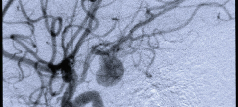
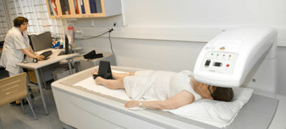
Volunteers underwent MRI or CT scans in order to get slice images of the human body. These images were then used to create a 3D model of the skeletal system. The models originally developed for machines were not applicable as such to humans due to the more complex geometry of the skeletal system.
“But complex geometry poses no problems for the powerful computers we have today,” explains Mikkola. “As for the muscular system, we relied on the expertise of our partners in Jyväskylä because we had no knowledge of large muscle groups and how they work.”
The human control system – the connections from the brain to the limbs – proved a tough simulation challenge: steering and control mechanisms are far more complex in humans than they are in machines. Even children spend at least a year learning how to walk.
The first models to simulate human walking proved unsuccessful, but the LUT team didn’t give up:
They solved the challenge by measuring the movements and the simple exercises performed by the participants in the experiments, tracking the movement of small points of measurement in 3D space and then programming the simulation model to repeat these movements. In other words, the simulation model was taught to repeat the exercise.
Impact the bones
Once the exercise was complete the researchers were able to move on to test and study the impact of the exercises on bone strain and bone mass building.
Simulation can provide information that can otherwise only be obtained by using so-called in vivo strain gauges attached to the bone surface. Strain gauge measurement involves invasive surgery and consequently poses a risk of infection, and for ethical reasons it would probably not even be considered in Finland, says Mikkola.
The team studied human walking in order to verify the technique to a level of accuracy that can produce meaningful results. They also had access to previous gauge data on walking, which provided a useful point of reference.
“The important thing is that nobody is harmed in the process of simulation,” he says. “We can also study bones under the surface, whereas strain gauges attached to the bone only yield information from a single point of the bone. We’re particularly interested in the hip area and the stresses it goes through when running.”
The researchers are hoping to gain a clearer view of how different exercises impact bone mass strengthening. The results could help in the development of exercise equipment that is ideally suited to strengthening the bones in the hip area.
Jumping and bouncing have beneficial effects
There are great variations in effect from one exercise to another:
“The leg press, for instance, is much more effective in terms of strengthening bone mass than the squat exercise because it induces more strains.”
Any kind of bouncing movement has beneficial effects in terms of strengthening the skeletal system; however, moderation is crucial:
“Exercises that involve intense stresses such as the triple jump are particularly effective, but triple jumpers tend to suffer from other complaints, such as degenerative arthritis.”
Tracing the causes of degenerative arthritis
A new research project is now underway to explore the aetiology of degenerative arthritis.
Degenerative arthritis is the most common type of arthritis, but its causes remain unknown. Since it affects older people in particular, it is expected that the problem will continue to grow in line with the ageing of the population. In Finland alone, the number of sufferers is estimated at around 400,000 (out of a total population of 5.4 million).
The project aims to bring relief for many of these people by shedding light on the aetiology of degenerative arthritis, providing guidance on prevention and by developing preventive equipment and safety systems.
This research will again require innovative approaches:
“The knee joint is highly complex in a mechanical sense. There are materials in the knee that are unknown to mechanical engineering,” says Mikkola.
The cartilage in the knee functions in rather the same way as sliding bearings in heavy duty machines. The problem is that as the cartilage wears away, it is lost forever. The only way to repair degenerative arthritis is to implant an artificial joint, but this is an extreme procedure that is avoided as far as possible, he explains.
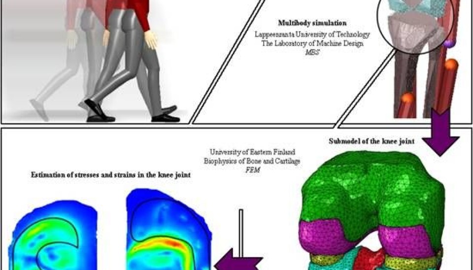
“We’re aiming to uncover the mechanisms of cartilage wear. In the first phase, our colleagues in Jyväskylä will again take measurements on human subjects, and then the simulation models we develop in Lappeenranta will be taught to repeat certain exercises.”
Once the exercise is complete, the information on the contact forces impacting the knee joint will be assessed by a highly detailed model to determine whether the contacts, forces and tensions are too high and possibly responsible for causing degenerative arthritis.
“We hope the new knowledge will pave the way towards developing new preventive and protective equipment, new exercises and better artificial joints,” Mikkola concludes.
Scientific links
- A full body musculoskeletal model based on flexible multibody simulation approach utilised in bone strain analysis during human locomotion
- Flexible Multibody Simulation Approach in the Dynamic Analysis of Bone Strains Activity