New invention looks deep into the soul of cancer cells
Danish scientists have invented a method that makes MR scanners up to 100,000 times more sensitive. This makes it possible to tell instantly whether a cancer treatment is effective.
When treating cancer patients, doctors need to know as soon as possible if a drug is working, or if it is necessary to try another form of treatment.
Today, doctors are forced to wait until it is possible to tell from the size of the tumour whether the medicine has had the desired effect. As this often takes a long time, the patient’s condition may have deteriorated in the meantime.
However, thanks to a new Danish invention it is now possible to follow the drug’s way into the cancer cells ‘live’, thus dramatically increasing the patient’s chance of receiving the right treatment from day one.
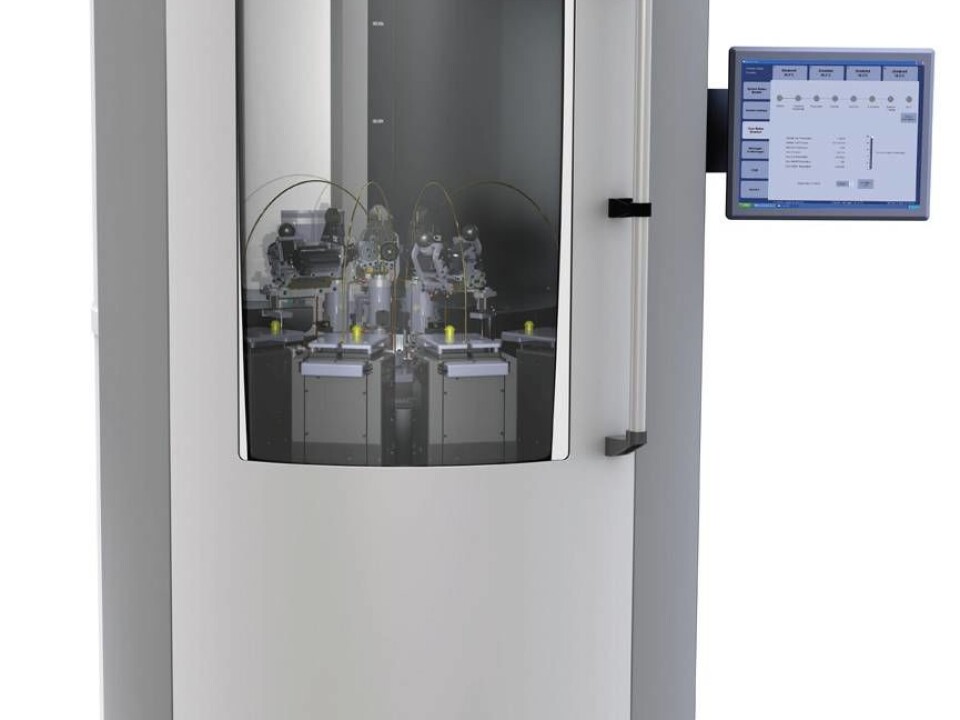
The new invention is a machine that can magnetise trace compounds. In combination with an MR scanner, it offers an image of hitherto unseen accuracy of what is going on inside the body and the cancer cells.

“We’re currently testing magnetised trace compounds in patients with prostate cancer, but we have already had success testing the method on animals. If the project succeeds, it will be a game changer in the battle against cancer,” says Jan Henrik Ardenkjær-Larsen, a professor of electrical engineering at the Technical University of Denmark (DTU).
Machine magnetises trace compounds
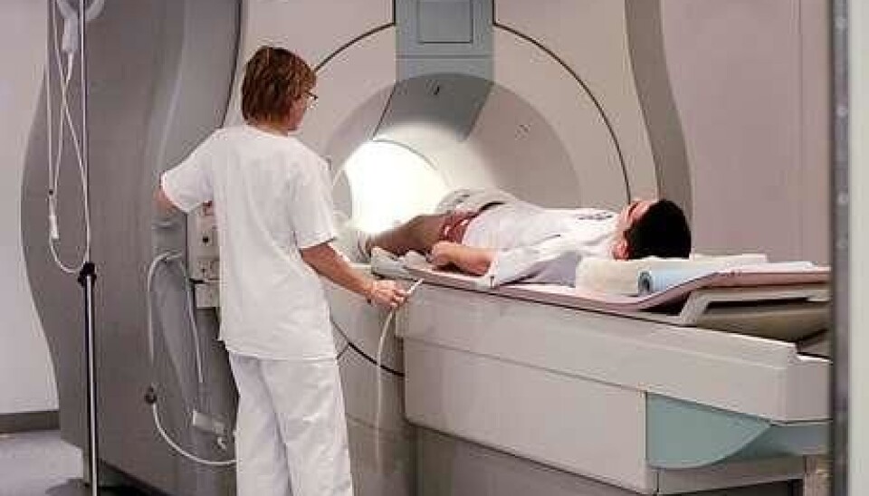
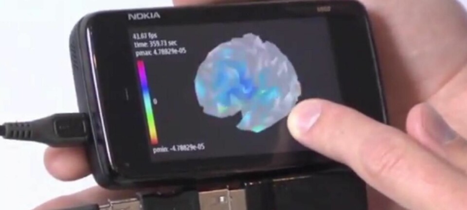
The background for the new invention is the MR scanner, whose functions include locating cancer cells in the body and assessing brain function.
MR scanners, however, have their limitations. It only gives a relatively grainy image of the tiny details that reveal the extent to which a cancer cell is responding to treatment.
With the new invention, researchers have set out to change this.
We have reason to believe that we will very soon be able to tell very early on in the process how cancer patients will react to treatment.
By magnetising compounds that are then absorbed by the cancer cells, Ardenkjær-Larsen and his colleagues can follow the compounds through the MR scanner and see whether it enters the cancer cells. Presumably, it can also be used to follow a cancer drug’s path into the body, and to see whether the cells absorb it.
“MR scanning is extremely useful for seeing organs, tumours and other aspects of anatomy. Unfortunately, it’s not very sensitive,” says Ardenkjær-Larsen.
“If we want to see how the biochemical processes in the body function, we therefore have to increase the sensitivity of the scanner. We achieve that by using a contrast medium that makes the MR scanner close to 100,000 times more sensitive.”
The contrast fluids are magnetised biological molecules like sugars, which are rarely harmful to people.
Prostate cancer detected in medical trials
The scientists behind the new invention are currently testing the magnetised trace compounds on patients with prostate cancer.
Cancer of the prostate gland is often in a dormant state, making it harmless. However, the cancer may become aggressive, making the patient’s health deteriorate rapidly.
Doctors have long needed new techniques that allow them to assess whether the cancer is aggressive or dormant. The new invention will make this possible.
By creating the trace compounds from biological molecules, the scientists can tell how quickly the cancer cells absorb the molecules. The faster the absorption occurs, the more aggressive the cancer is. Scientists can also magnetise cancer drugs and see if the cancer cells absorb those.
“In our experiments, we saw that the tumours lit up when we gave the patients the magnetised trace compounds. In some cases, we also discovered new tumours that hadn’t previously been detected,” he says.
The new invention magnetises molecules
Ardenkjær-Larsen’s new machine plays a key role in the efforts to figure out what goes on inside the cancer cells.
The machine is capable of magnetising trace compounds. This means that the trace compounds become more visible when captured by the MR scanner.
“It’s pure magnetism. If you’re trying to achieve a more precise image in an MR scan, you need a more powerful magnet to make the magnetic orientation of the molecules conform and give a signal,” he says.
“We have reason to believe that we will very soon be able to tell very early on in the process how cancer patients will react to treatment.”
Ardenkjær-Larsen isn’t alone in believing that the machine will revolutionise the battle against cancer.
“We are lucky to have someone like Jan Henrik Ardenkjær-Larsen, who has invented a method to hyperpolarise trace compounds and make the signal from the MR scanners thousands of times stronger,” says Professor Hans Stødkilde Jørgensen, who carries out research in the use of MR scanners at Skejby Hospital in Denmark.
“At the moment, we’re trying to get permission to use this type of machine. The invention is comparable to putting an amplifier on an indoor aerial, allowing it to receive thousands more TV signals. In this case, it’s signals that tell us something about the body.”
--------------------------------
Read the Danish version of this article at videnskab.dk
Translated by: Iben Gøtzsche Thiele
Scientific links
- Increase in signal-to-noise ratio of > 10,000 times in liquid-state NMR, PNAS, doi: 10.1073/pnas.1733835100
- Detecting tumor response to treatment using hyperpolarized 13C magnetic resonance imaging and spectroscopy, Nature, doi:10.1038/nm1650


