Digital morgue to help solve crimes
Digital 3D animation of dead bodies aims to revolutionise police investigation and improve accuracy in crime scene reconstructions.
We are at the Department of Forensic Medicine at the University of Copenhagen – far from Hollywood and the forensic glamour we know from US crime shows like CSI, Bones and Numbers.
This will, however, soon become the scene of the development of a brand new forensic procedure that would fit in well in a US crime show.
Here, Italian scientist Chiara Villa is working on creating 3D animations of deceased crime victims in an effort to make more accurate crime scene reconstructions, ultimately helping forensic scientists solve crimes.
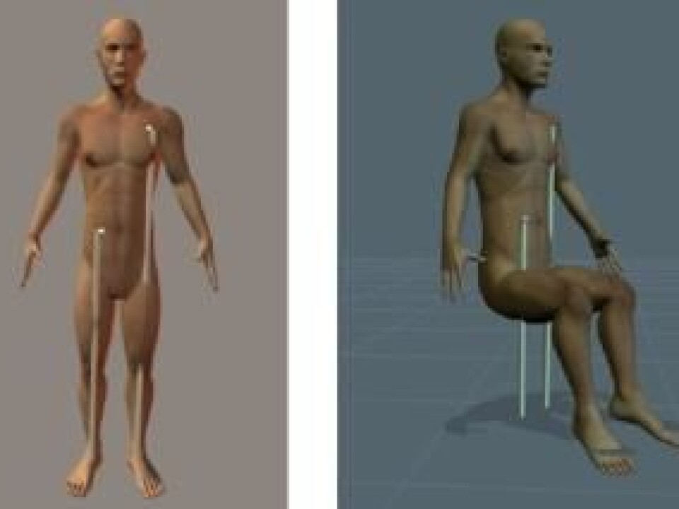
“The idea is to develop a virtual 3D model of the dead body, which can then be animated. This makes it possible to reconstruct body positions and thus improve the accuracy of forensic examinations,” says Villa.
Digital crime reconstruction

The study forms part of Villa’s postdoctoral project at the University of Copenhagen, titled ‘Walking dead: Merging modern imaging and 3D visualization techniques to provide a virtual reality support for forensic pathology practice’.
”By creating a 3D model, it will be possible to recreate the exact body position at the time of the crime. With existing methods it is only possible to reach approximate results using a dummy.”
This enables forensic pathologists and police investigators to determine with very little uncertainty the position and motion pathways of the victim when the shot hits. This is crucial not only when the police need to identify a perpetrator, but also in the subsequent trial.
”We hope the method will be reliable enough to be used as evidence in court. It would give a much more realistic picture of the incident,” says Villa.
Virtual archiving of corpses
The digital nature of the new method opens up for yet another important function. Crime cases often take a long time to solve, and it can take months, even years, before the conclusive evidence suddenly appears.
By then, the physical evidence – the dead body – may have decayed or disappeared altogether. This can lead to inaccuracies in the production of evidence.
”With 3D visualisations we can build up a digital archive, which allows us to analyse saved data based on the latest evidence. This allows us to recreate the sequence of events with more accuracy.”
Digital evidence is independent of time, and you will always have a digital copy of ’the corpse’ stored on a computer, so you can test the new evidence – much like a digitalised morgue.
Scans, X-rays, lasers and computer gaming technology
The technology required for developing a 3D model of dead bodies has already been invented. This means that Villa will not need to develop a new technology, but rather combine or integrate data from a variety of technologies to create the virtual 3D model, which will consist of an external as well as an internal visualisation of the corpse.
“By using CT and MRI technology, we can visualise the inner part of the body, but not the outer part in very great detail. I will be using photogrammetry and laser scanning to accurately recreate the outer part of the body.”
Villa’s postdoc study does not start until next month, so the method of merging the various technologies has not yet been finalised. The overall plan, however, is to use software to compile and organise the data from the various technologies.
This software includes the program Poser, which is used to animate real persons for computer games, such as when Angelina Jolie was modelled for the Tomb Raider game.
Copenhagen as a forensic nerve centre
Chiara Villa is born and bred in Italy, but went to Denmark to do her research because Copenhagen is a nerve centre when it comes to this specific field within forensic medicine.
“I was educated in Milan and did my research there until I moved to Denmark. There are not many places in the world where forensic departments use both CT and MRI scanners,” she says, adding that the ‘well-equipped’ department is what prompted her to move to Copenhagen.
In addition to Copenhagen, CT and MRI technology is also being combined in Switzerland, Australia and probably also in the US, she says. This is, however, an area in flux, and she believes that her method will be in demand once it is completed a couple of years from now:
”I hope the method will be ready to use immediately. For the departments where the technologies have already been installed, there should be no problems with starting to use the method immediately.”
------------------
Read the Danish version of this article at videnskab.dk
